9GPB

THE ALLOSTERIC TRANSITION OF GLYCOGEN PHOSPHORYLASE


Experimental Data Snapshot

  • Method: X-RAY DIFFRACTION
  • Resolution: 2.90 Å
  • R-Value Work: 
    0.177 (Depositor) 

wwPDB Validation   3D Report Full Report


This is version 2.0 of the entry. See complete history


Literature

The allosteric transition of glycogen phosphorylase.

Barford, D.Johnson, L.N.

(1989) Nature 340: 609-616

  • DOI: https://doi.org/10.1038/340609a0
  • Primary Citation of Related Structures:  
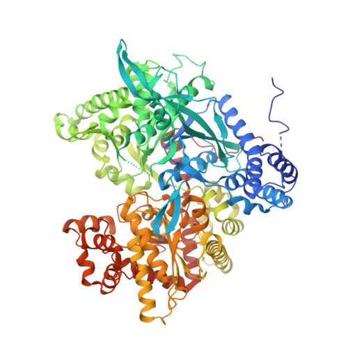
    9GPB

  • PubMed Abstract: 

    The crystal structure of R-state glycogen phosphorylase b has been determined at 2.9 A resolution. A comparison of T-state and R-state structures of the enzyme explains its cooperative behaviour on ligand binding and the allosteric regulation of its activity. Communication between catalytic sites of the dimer is provided by a change in packing geometry of two helices linking each site with the subunit interface. Activation by AMP or by phosphorylation results in a quaternary conformational change that switches these two helices into the R-state conformation.


  • Organizational Affiliation

    Laboratory of Molecular Biophysics, University of Oxford, UK.


Macromolecules
Find similar proteins by:  (by identity cutoff)  |  3D Structure
Entity ID: 1
MoleculeChains Sequence LengthOrganismDetailsImage
Glycogen phosphorylase, muscle form
A, B, C, D
842Oryctolagus cuniculusMutation(s): 0 
Gene Names: PYGM
EC: 2.4.1.1
UniProt
Find proteins for P00489 (Oryctolagus cuniculus)
Explore P00489 
Go to UniProtKB:  P00489
Entity Groups  
Sequence Clusters30% Identity50% Identity70% Identity90% Identity95% Identity100% Identity
UniProt GroupP00489
Sequence Annotations
Expand
  • Reference Sequence
Small Molecules
Ligands 1 Unique
IDChains Name / Formula / InChI Key2D Diagram3D Interactions
SO4
Query on SO4

Download Ideal Coordinates CCD File 
E [auth A]
F [auth A]
G [auth A]
H [auth B]
I [auth B]
E [auth A],
F [auth A],
G [auth A],
H [auth B],
I [auth B],
J [auth B],
K [auth C],
L [auth C],
M [auth C],
N [auth D],
O [auth D],
P [auth D]
SULFATE ION
O4 S
QAOWNCQODCNURD-UHFFFAOYSA-L
Modified Residues  1 Unique
IDChains TypeFormula2D DiagramParent
LLP
Query on LLP
A, B, C, D
L-PEPTIDE LINKINGC14 H22 N3 O7 PLYS
Experimental Data & Validation

Experimental Data

  • Method: X-RAY DIFFRACTION
  • Resolution: 2.90 Å
  • R-Value Work:  0.177 (Depositor) 
Space Group: P 1 21 1
Unit Cell:
Length ( Å )Angle ( ˚ )
a = 119α = 90
b = 190β = 109.35
c = 88.2γ = 90
Software Package:
Software NamePurpose
X-PLORrefinement

Structure Validation

View Full Validation Report


Entry History 

Deposition Data

Revision History  (Full details and data files)

  • Version 1.0: 1992-10-15
    Type: Initial release
  • Version 1.1: 2008-03-25
    Changes: Version format compliance
  • Version 1.2: 2011-07-13
    Changes: Non-polymer description, Version format compliance
  • Version 1.3: 2017-11-29
    Changes: Derived calculations, Other
  • Version 2.0: 2024-12-25
    Changes: Advisory, Atomic model, Data collection, Database references, Derived calculations, Non-polymer description, Polymer sequence, Source and taxonomy, Structure summary