


News
Visualize in 3D with PV
05/26
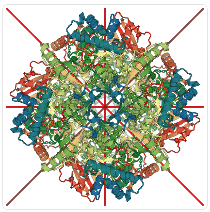 PV view of rubisco (PDB ID 4F0H).
An experimental version of the Protein Viewer (PV) has been added to the visualization options offered by the RCSB PDB for proteins and nucleic acid structures on each Structure Summary page. PV uses WebGL and enables hardware-accelerated graphics in modern web and mobile browsers.
PV can display symmetric structures aligned along the symmetry axes. It can also be launched in full screen or standalone windows.
For more information on PV, please visit http://biasmv.github.io/pv/.
