


News
Visualize Structure Quality Metrics in 3D
11/22
wwPDB Validation Reports are available for every entry to provide an assessment of the quality of a structure and highlight specific concerns by considering the model coordinates, experimental data, and fit between the two.
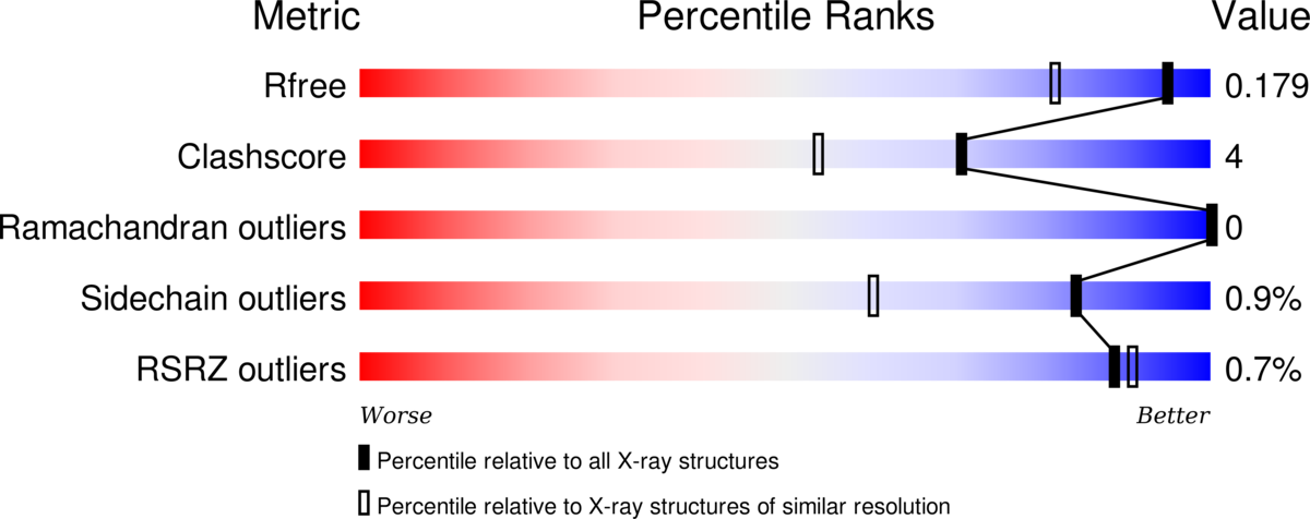
RCSB PDB Structure Summary pages contain the “slider” graphic from these reports that provides a visual summary and also link to the full PDF report.
New options in the NGL viewer map wwPDB Validation Report information onto the 3D structure. Clashes can be displayed as pink disks, and the full structure can be shown using “Geometry Quality” and “Density Fit” coloring schemes. More detailed information about this feature is available.
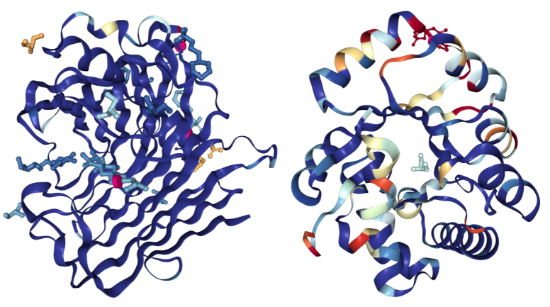 Color By Density Fit: Hydrolases colored using the “Density Fit” scheme.
 On the left is a structure of Endoglucanase A (PDB ID 3WY6) with a generally good fit; on the right a structure of Ribonuclease P protein component 3 (PDB ID 3WYZ), which has areas of more problematic fit. For details regarding RSRZ and RSCC, please consult the Validation Report User Guide for X-ray structures.
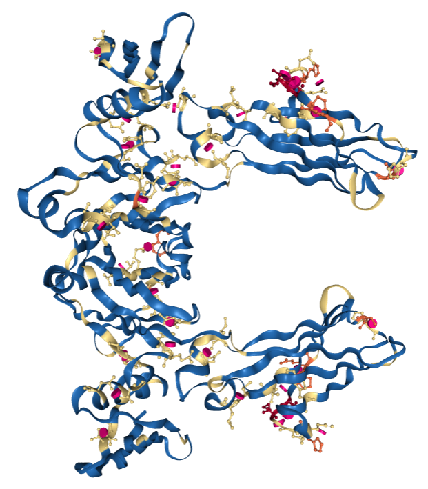 Color By Geometry Quality: PDB 1FCC, a 3.2Å resolution structure with worse overall quality relative to all X-ray structures colored by Geometry Quality.
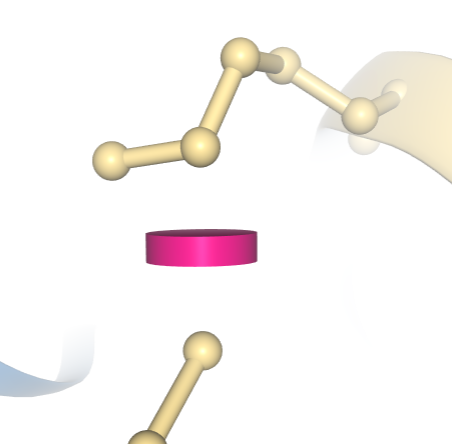 Display Clashes Between Atoms: A clash between two atoms in PDB 1D66 is indicated by a pink disc, showing how much the atoms’ vdW spheres overlap. In this example, the structure has been colored using the “Geometry Quality” scheme, which indicates that this clash is the only geometric issue for these two residues.

